baby chest x ray exposure
Are Chest X Rays Safe For Babies. Radiation exposure from X-rays may slightly raise the risk of later cancer especially in children who have had many tests with high radiation exposure.

Antarctica Neo Archive Que X Ray C Spine Ap View Image File No 0305 X Ray View Image History Of Science
All distal extremity exposures are taken at 110115 cm SID.

. Canada uses lower kV Figure 1 but Norway uses lower mAs Figure 2. In such cases the radiation dose to the conceptus and subsequent risks should be estimated. Radiation exposure from X-rays does not pose any short-term problems.
The chest X-ray is the most frequently ordered radiological. Exposure 527 Neonatal Chest X-ray. As a benchmark for other medical imaging departments and to promote discussion on digital X.
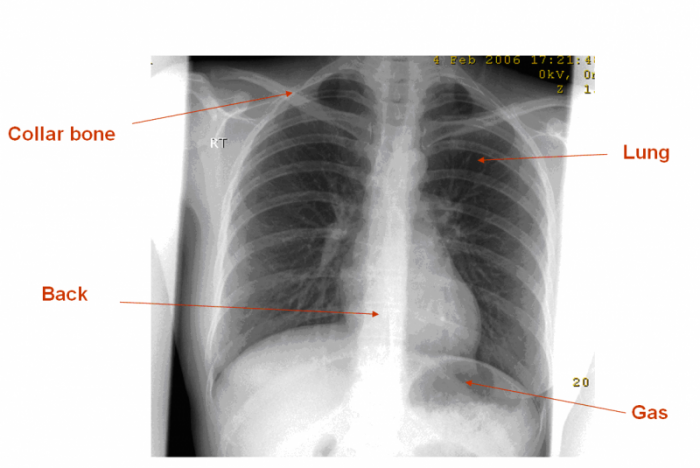
The publication of this study and exposure chart could act. In addition to radiation exposure Burstein points out theres a high association between receiving a chest X-ray for bronchiolitis and antibiotic prescribing which is. The left hemidiaphragm should be visible to the edge of the spine.
It might look scary but it. The risk of harm to your baby depends on your babys gestational age and the amount of radiation exposure. Lateral cervical spines are taken at 150 cm.
RESULTS AND DISCUSSION Table 1 shows a summary of annual percentage frequencies of paediatric examinations within each age group each column sums to 100. Its use is to immobilize the child so that they dont wiggle around. Your doctor or the hospital will run tests including blood sugar levels blood tests heart tests a urine test to rule.
The chin should not be superimposing any structures. Most researchers agree that babies who receive a small dose of radiation equal to 500 chest x -rays or less at any time during pregnancy do not have an increased risk for birth defects. The only increased risk to these babies is a slightly higher chance of having cancer later in life less than 2 higher than the.
However these dose levels arent used in diagnostic imaging. The entire lung fields should be visible from the apices down to the lateral costophrenic angles. Because they spin around the body taking multiple images CT scans can deliver radiation doses that are up to 200 times higher than an average chest X.
All distal extremity exposures are taken at 110115 cm SID. She had scanning yesterday and it is said that baby is 6 weeks 3 days old. Erect chest X-rays are taken at 180 cm.
See Safety in X-ray Interventional Radiology and Nuclear Medicine Procedures for more information. ESD was determined using DoseCal software and patients exposure factors X-ray tube output and backscatter factors BSFs in accordance with the following formula Davies et al 1997. Long-term problems are very small.
X-ray exams provide valuable information about your health and help your doctor make an accurate diagnosis. The doctor may order an X-ray to understand the extent of the babys injuries. Exposure to high-dose radiation two to eight weeks after conception might.
Erect chest X-rays are taken at 180 cm. Erect chest X-rays are taken at 180 cm. Although X-rays are still occasionally over or under exposed a discussion of penetration now best serves as a reminder to check behind the heart.
Radiation exposure is known to damage the cells that were exposed and can lead to cancer. A well penetrated chest X-ray is one where the vertebrae are just visible behind the heart. The purpose of this study was the calculation and presentation of fetal dose and subsequent risks resulted from different X-ray examinations.
It is wise to be concerned about radiation exposure but the amount of radiation in a chest x-ray is very small. Medical exposure 5 and published in UNSCEAR-2000 Report 6. Pregnant women are sometimes exposed to ionizing radiation in radiology examinations for various reasons.
Not providing the proper protection during the X-ray or over-radiating the baby can cause serious harm. Exposure to extremely high-dose radiation in the first two weeks after conception might result in a miscarriage. Most neonatal chest X-rays are AP films unless the baby is made to lie prone Lucency of soft tissue shadow - darker the soft tissue more.
Lateral cervical spines are taken at 150 cm. Often smaller mini detectors are used for the neonate chest x-ray. Full legfull spine imaging is performed at 180 cm using CR.
Your doctor may use x-rays to help place tubes or other devices in your body or to treat disease. The median exposure parameters in Canada for mobile neonatal chest imaging are 60 kV at 15 mAs with an inherent filtration of 20 mm Al Figure 3. Arms are not superimposed over lateral chest wall this can mimic pleural thickening.
Background exposure groups based on patient size. Bronchopulmonary dysplasia bpd sometimes called chronic lung disease is a problem with how a babys lung tissue develops. The most sensitive time period for central nervous system teratogenesis is between.
I use one every time I do a chest X-ray on a baby. 35 cm x 43 cm or 43 cm x 35 cm. For example the amount of exposure to the fetus from a two-view chest x-ray of the mother is only 000007 rad.
There are also differences in exposure factors between the two countries. Baby Chest X Ray Exposure. It is quite obvious that chest examination is by far the most frequently used diagnostic X-ray imaging procedure and accounts.
Full legfull spine imaging is performed at 180 cm using CR. Rotation Soft tissue bone Thymus -.

Radiologic Technique Charts Tech Nique Chart For Techniquetechnique Charts Radiologic Radiology Schools Radiology Student Radiology Technician

X Ray Imaging For Covid 19 Patients

Neonate Chest Supine View Radiology Reference Article Radiopaedia Org

Are Children Being Exposed To Excessive X Ray Radiation During Chest X Rays Doctor For Kids Children Parenting Hacks

Chest X Ray Basic Interpretation X Ray Medical Terminology Study Interpretation

Pediatric Chest Supine View Radiology Reference Article Radiopaedia Org

X Ray Imaging For Covid 19 Patients

Pem Pearls Chest Radiographs For Shortness Of Breath

Approach To The Chest X Ray Cxr Undergraduate Diagnostic Imaging Fundamentals

Antarctica Neo Archive Que X Ray Chest Pa View Image File No 0005 X Ray View Image History Of Science
Ap T Spine Image Radiology Technologist Radiologic Technology Radiography

What Is An X Ray For Kids Radiology And Medical Imaging
Plain Radiograph X Ray Insideradiology

27 Crazy Images Of Medical Treatments Through History Medical Photography Radiology Humor Medical History

Assessment Of Cxr Positioning Views How To Read A Chest X Ray Part 4 Medzcool Youtube

Pediatric Chest X Ray In Covid 19 Infection European Journal Of Radiology

Digital X Ray Centre In Ahmedabad X Ray Radiation Dose Electromagnetic Radiation

Good Lateral Elbow Diagnostic Imaging Radiology Schools Medical Mnemonics
What To Look For On A Chest X Ray Slideshow